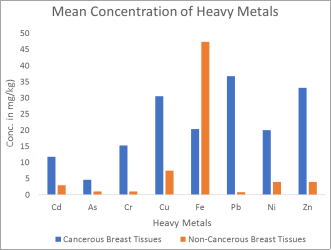
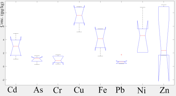
Breast cancer is prevalent in northern Nigerian women most especially Jos, Plateau State owing to anthropogenic activities such as solid earth mineral mining. In this study, Atomic Absorption Spectrometry was used to determine the levels of eight heavy metals (Cd, As, Cr, Cu, Fe, Pb, Ni and Zn) in cancerous and non-cancerous breast tissues of Jos Nigerian Women. The concentration of heavy metals ranged from 1.08 to 29.34 mg/kg, 0.29 to 10.76 mg/kg, 0.35 to 51.93 mg/kg, 5.15 to 62.93 mg/kg, 11.64 to 51.10 mg/kg, 0.42 to 83.16 mg/kg, 2.08 to 43.07 mg/kg and 1.67 to 71.53 mg/kg for Cd, As, Cr, Cu, Fe, Pb, Ni and Zn respectively. Using MATLAB R2016a, significant differences (tv = 0.0041 – 0.0317) existed between the levels of all the heavy metals in cancerous and non-cancerous breast tissues except Fe. At 0.01 level of significance, positive significant correlation existed between Pb and Fe, Pb and Cu, Pb and Fe, Ni and Fe, Cr and Pb, as well as Ni and Cr (r = 0.583 – 0.998) in cancerous breast tissues. Using ANOVA, significant differences also occurred in the levels of these heavy metals in cancerous breast tissues (p = 1.910510×10⁻²⁶). The relatively high levels of the cancer-induced heavy metals (Cd, As, Cr and Pb) compared with control indicated contamination or exposure to heavy metals which could be the major cause of cancer in these female subjects.
| Published in | World Journal of Public Health (Volume 9, Issue 2) |
| DOI | 10.11648/j.wjph.20240902.18 |
| Page(s) | 186-193 |
| Creative Commons |
This is an Open Access article, distributed under the terms of the Creative Commons Attribution 4.0 International License (http://creativecommons.org/licenses/by/4.0/), which permits unrestricted use, distribution and reproduction in any medium or format, provided the original work is properly cited. |
| Copyright |
Copyright © The Author(s), 2024. Published by Science Publishing Group |
Atomic Absorption Spectroscopy, Breast Cancer, Heavy Metal Exposure
Elements | Cancerous breast tissues (n = 25) | Non-cancerous breast tissues (n = 6) | ||
|---|---|---|---|---|
Range (mg/kg) | Mean ± SD | Range (mg/kg) | Mean ± SD | |
Cd | 4.02 - 29.34 | 11.75 ± 1.22 | 1.08 - 4.92 | 2.94 ± 0.58 |
As | 0.75 - 10.76 | 4.67 ± 0.50 | 0.29 -1.60 | 1.05 ± 0.19 |
Cr | 1.92 - 51.93 | 15.28 ± 2.61 | 0.35 - 1.75 | 0.99 ± 0.22 |
Cu | 17.06 - 62.93 | 30.53 ± 2.39 | 5.15 - 8.93 | 7.45 ± 0.66 |
Fe | 11.64 - 42.22 | 20.39 ± 1.63 | 38.10 - 51.10 | 47.27 ± 6.00 |
Pb | 22.85 - 83.16 | 38.68 ± 3.24 | 0.42 - 1.74 | 0.78 ± 0.20 |
Ni | 11.85 - 43.07 | 20.04 ± 1.67 | 2.08 - 5.61 | 3.93 ± 0.66 |
Zn | 19.54 - 71.53 | 33.12 ± 2.75 | 1.67 - 9.11 | 3.92 ± 1.30 |
Element | t-value | Remark |
|---|---|---|
Cd | 0.0307 | S |
As | 0.0273 | S |
Cr | 0.0063 | S |
Cu | 0.0273 | S |
Fe | 0.0571 | NS |
Pb | 0.0317 | S |
Ni | 0.0041 | S |
Zn | 0.0139 | S |
Elements | Concentration of cancerous breast tissues in this study (mg/kg) | Sarita (2012) | Mohammadi et al (2014) | Pasha et al (2008) | Mehmet et al (2007) |
|---|---|---|---|---|---|
Cd | 11.75 | 38.66 | 2.64 | 33.00 | |
As | 4.67 | ||||
Cr | 15.28 | 10.40 | |||
Cu | 30.53 | 32.30 | 4.30 | ||
Fe | 20.39 | 42.00 | |||
Pb | 36.68 | 35.69 | 13.20 | ||
Ni | 20.04 | ||||
Zn | 33.12 | 13.00 | 45.00 |
Cd | As | Cr | Cu | Fe | Pb | Ni | Zn | |
|---|---|---|---|---|---|---|---|---|
Cd | 1.000 | 0.583** | 0.442 | 0.337 | 0.281 | 0.287 | 0.288 | 0.275 |
As | 0.583** | 1.000 | 0.071 | -0.004 | -0.012 | -0.011 | -0.090 | -0.020 |
Cr | 0.442* | 0.070 | 1.000 | 0.799** | 0.789** | 0.788** | 0.787** | 0.782** |
Cu | 0.337 | -0.004 | 0.799** | 1.000 | 0.982** | 0.981** | 0.981** | 0.979** |
Fe | 0.281 | -0.012 | 0.789** | 0.982** | 1.000 | 0.998** | 0.998** | 0.997** |
Pb | 0.287 | -0.110 | 0.288** | 0.982** | 0.998** | 1.000** | 1.000** | 1.000** |
Ni | 0.288 | -0.009 | 0.787** | 0.981** | 0.998** | 1.000** | 1.000 | 1.000** |
Zn | 0.275 | -0.020 | 0.782** | 0.979** | 0.997** | 1.000** | 1.000** | 1.000 |
IDC | Invasive Ductal Carcinoma |
ILC | Invasive Lobular Carcinoma |
IBC | Inflammatory Breast Cancer |
AAS | Atomic Absorption Spectrometer |
| [1] | Angell, B., Sanuade, O., Adetifa, I. M. O., Okeke, I. N., Adamu, A. L., Aliyu, M. H., Ameh, E. A., Kyari, F., Gadanya, M. A., Mabayoje, D. A., Yinka-Ogunleye, A., Oni, T., Jalo, R. I., Tsiga-Ahmed, F. I., Dalglish, S. L., Abimbola, S., Colbourn, T., Onwujekwe, O., Owoaje, E. T., … Abubakar, I. (2022). Population health outcomes in Nigeria compared with other west African countries, 1998–2019: a systematic analysis for the Global Burden of Disease Study. Lancet (London, England), 399(10330), 1117. |
| [2] | Azubuike, S. O., Muirhead, C., Hayes, L., & McNally, R. (2018). Rising global burden of breast cancer: The case of sub-Saharan Africa (with emphasis on Nigeria) and implications for regional development: A review. World Journal of Surgical Oncology, 16(1), 1–13. |
| [3] | Batyrova, G., Kononets, V., Amanzholkyzy, A., Tlegenova, Z., & Umarova, G. (2022). Chromium as a Risk Factor for Breast Cancer: A Meta-Analysis. Asian Pacific Journal of Cancer Prevention: APJCP, 23(12), 3993. |
| [4] | Brown, R. A. M., Richardson, K. L., Kabir, T. D., Trinder, D., Ganss, R., & Leedman, P. J. (2020). Altered Iron Metabolism and Impact in Cancer Biology, Metastasis, and Immunology. Frontiers in Oncology, 10. |
| [5] | Büsselberg, D., & Florea, A. M. (2011). Metals and Breast Cancer: Risk Factors or Healing Agents? Journal of Toxicology, 2011. |
| [6] | Buxton, S., Garman, E., Heim, K. E., Lyons-Darden, T., Schlekat, C. E., Taylor, M. D., & Oller, A. R. (2019). Concise Review of Nickel Human Health Toxicology and Ecotoxicology. Inorganics 2019, Vol. 7, Page 89, 7(7), 89. |
| [7] | Fatiregun, O. A., Oluokun, T., Lasebikan, N. N., Nwachukwu, E., Ibraheem, A. A., & Olopade, O. (2021). Breast Cancer Research to Support Evidence-Based Medicine in Nigeria: A Review of the Literature. JCO Global Oncology, 7, 384–390. |
| [8] | Feng, Y., Spezia, M., Huang, S., Yuan, C., Zeng, Z., Zhang, L., Ji, X., Liu, W., Huang, B., Luo, W., Liu, B., Lei, Y., Du, S., Vuppalapati, A., Luu, H. H., Haydon, R. C., He, T. C., & Ren, G. (2018). Breast cancer development and progression: Risk factors, cancer stem cells, signaling pathways, genomics, and molecular pathogenesis. Genes & Diseases, 5(2), 77. |
| [9] | Khan, S., Shah, I. A., Muhammad, S., Malik, R. N., & Shah, M. T. (2015). Arsenic and Heavy Metal Concentrations in Drinking Water in Pakistan and Risk Assessment: A Case Study. Human and Ecological Risk Assessment, 21(4), 1020–1031. |
| [10] | Łukasiewicz, S., Czeczelewski, M., Forma, A., Baj, J., Sitarz, R., & Stanisławek, A. (2021). Breast Cancer—Epidemiology, Risk Factors, Classification, Prognostic Markers, and Current Treatment Strategies—An Updated Review. Cancers, 13(17). |
| [11] | Manz, D. H., Blanchette, N. L., Paul, B. T., Torti, F. M., & Torti, S. V. (2016). Iron and cancer: recent insights. Annals of the New York Academy of Sciences, 1368(1), 149. |
| [12] | Nishida, N., Yano, H., Nishida, T., Kamura, T., & Kojiro, M. (2006). Angiogenesis in Cancer. Vascular Health and Risk Management, 2(3), 213. |
| [13] | Richter, P., Faroon, O., & Pappas, R. S. (2017). Cadmium and Cadmium/Zinc Ratios and Tobacco-Related Morbidities. International Journal of Environmental Research and Public Health, 14(10). |
| [14] | Semu, E. (2019). Heavy Metals and Organopesticides: Ecotoxicology, Health Effects and Mitigation Options with Emphasis on Sub-Saharan Africa. Toxicology: Current Research, 3(1), 1–14. |
| [15] | Shah, N. R., & Wong, T. (2006). Current breast cancer risks of hormone replacement therapy in postmenopausal women. Expert Opinion on Pharmacotherapy, 7(18), 2455. |
| [16] | Tchounwou, P. B., Yedjou, C. G., Patlolla, A. K., & Sutton, D. J. (2012). Heavy metal toxicity and the environment. EXS, 101, 133–164. |
| [17] | Umesh C. Gupta, & Subhas C. Gupta. (2016). Heavy Metal Toxicity in Humans and its Preventive and Control Measures. Current Nutrition & Food Science, 7(4), 221–231. |
| [18] | Xiao, Y., Ma, D., Ruan, M., Zhao, S., Liu, X. Y., Jiang, Y. Z., & Shao, Z. M. (2017). Mixed invasive ductal and lobular carcinoma has distinct clinical features and predicts worse prognosis when stratified by estrogen receptor status. Scientific Reports, 7(1). |
| [19] | Yue, W., Wang, J. P., Li, Y., Fan, P., Liu, G., Zhang, N., Conaway, M., Wang, H., Korach, K. S., Bocchinfuso, W., & Santen, R. (2010). Effects of estrogen on breast cancer development: role of estrogen receptor independent mechanisms. International Journal of Cancer. Journal International Du Cancer, 127(8), 1748. |
| [20] | Zheng, Y., Walsh, T., Gulsuner, S., Casadei, S., Lee, M. K., Ogundiran, T. O., Ademola, A., Falusi, A. G., Adebamowo, C. A., Oluwasola, A. O., Adeoye, A., Odetunde, A., Babalola, C. P., Ojengbede, O. A., Odedina, S., Anetor, I., Wang, S., Huo, D., Yoshimatsu, T. F., … Olopade, O. I. (2018). Inherited Breast Cancer in Nigerian Women. Journal of Clinical Oncology, 36(28), 2820. |
APA Style
Idowu, O. P., Oyedele, O. O., Oluwadare, O. J., Igboama, W. N., Dolapo, O. S., et al. (2024). Atomic Absorption Spectroscopic Analysis of Heavy Metals in Cancerous Breast Tissues Among Women in Jos, Nigeria. World Journal of Public Health, 9(2), 186-193. https://doi.org/10.11648/j.wjph.20240902.18
ACS Style
Idowu, O. P.; Oyedele, O. O.; Oluwadare, O. J.; Igboama, W. N.; Dolapo, O. S., et al. Atomic Absorption Spectroscopic Analysis of Heavy Metals in Cancerous Breast Tissues Among Women in Jos, Nigeria. World J. Public Health 2024, 9(2), 186-193. doi: 10.11648/j.wjph.20240902.18
AMA Style
Idowu OP, Oyedele OO, Oluwadare OJ, Igboama WN, Dolapo OS, et al. Atomic Absorption Spectroscopic Analysis of Heavy Metals in Cancerous Breast Tissues Among Women in Jos, Nigeria. World J Public Health. 2024;9(2):186-193. doi: 10.11648/j.wjph.20240902.18
@article{10.11648/j.wjph.20240902.18,
  author = {Opeyemi Peter Idowu and Oketayo Oyebamiji Oyedele and Oluwatimilehin Joshua Oluwadare and Wilfred Nwabueze Igboama and Olaniyan Suaib Dolapo and Lawan Ezekiel and Catherine Ignatius and Mashor Mbwas Isaac and Audu Ruth Danbaki and Adefemi Frank Olasele and Hamzat Toheeb Tunde and Bamidele Lateef and Akinnubi Rufus Temidayo},
  title = {Atomic Absorption Spectroscopic Analysis of Heavy Metals in Cancerous Breast Tissues Among Women in Jos, Nigeria
},
  journal = {World Journal of Public Health},
  volume = {9},
  number = {2},
  pages = {186-193},
  doi = {10.11648/j.wjph.20240902.18},
  url = {https://doi.org/10.11648/j.wjph.20240902.18},
  eprint = {https://article.sciencepublishinggroup.com/pdf/10.11648.j.wjph.20240902.18},
  abstract = {Breast cancer is prevalent in northern Nigerian women most especially Jos, Plateau State owing to anthropogenic activities such as solid earth mineral mining. In this study, Atomic Absorption Spectrometry was used to determine the levels of eight heavy metals (Cd, As, Cr, Cu, Fe, Pb, Ni and Zn) in cancerous and non-cancerous breast tissues of Jos Nigerian Women. The concentration of heavy metals ranged from 1.08 to 29.34 mg/kg, 0.29 to 10.76 mg/kg, 0.35 to 51.93 mg/kg, 5.15 to 62.93 mg/kg, 11.64 to 51.10 mg/kg, 0.42 to 83.16 mg/kg, 2.08 to 43.07 mg/kg and 1.67 to 71.53 mg/kg for Cd, As, Cr, Cu, Fe, Pb, Ni and Zn respectively. Using MATLAB R2016a, significant differences (tv = 0.0041 – 0.0317) existed between the levels of all the heavy metals in cancerous and non-cancerous breast tissues except Fe. At 0.01 level of significance, positive significant correlation existed between Pb and Fe, Pb and Cu, Pb and Fe, Ni and Fe, Cr and Pb, as well as Ni and Cr (r = 0.583 – 0.998) in cancerous breast tissues. Using ANOVA, significant differences also occurred in the levels of these heavy metals in cancerous breast tissues (p = 1.910510×10⁻²⁶). The relatively high levels of the cancer-induced heavy metals (Cd, As, Cr and Pb) compared with control indicated contamination or exposure to heavy metals which could be the major cause of cancer in these female subjects.
},
 year = {2024}
}
TY  - JOUR
T1  - Atomic Absorption Spectroscopic Analysis of Heavy Metals in Cancerous Breast Tissues Among Women in Jos, Nigeria

AU  - Opeyemi Peter Idowu
AU  - Oketayo Oyebamiji Oyedele
AU  - Oluwatimilehin Joshua Oluwadare
AU  - Wilfred Nwabueze Igboama
AU  - Olaniyan Suaib Dolapo
AU  - Lawan Ezekiel
AU  - Catherine Ignatius
AU  - Mashor Mbwas Isaac
AU  - Audu Ruth Danbaki
AU  - Adefemi Frank Olasele
AU  - Hamzat Toheeb Tunde
AU  - Bamidele Lateef
AU  - Akinnubi Rufus Temidayo
Y1  - 2024/05/24
PY  - 2024
N1  - https://doi.org/10.11648/j.wjph.20240902.18
DO  - 10.11648/j.wjph.20240902.18
T2  - World Journal of Public Health
JF  - World Journal of Public Health
JO  - World Journal of Public Health
SP  - 186
EP  - 193
PB  - Science Publishing Group
SN  - 2637-6059
UR  - https://doi.org/10.11648/j.wjph.20240902.18
AB  - Breast cancer is prevalent in northern Nigerian women most especially Jos, Plateau State owing to anthropogenic activities such as solid earth mineral mining. In this study, Atomic Absorption Spectrometry was used to determine the levels of eight heavy metals (Cd, As, Cr, Cu, Fe, Pb, Ni and Zn) in cancerous and non-cancerous breast tissues of Jos Nigerian Women. The concentration of heavy metals ranged from 1.08 to 29.34 mg/kg, 0.29 to 10.76 mg/kg, 0.35 to 51.93 mg/kg, 5.15 to 62.93 mg/kg, 11.64 to 51.10 mg/kg, 0.42 to 83.16 mg/kg, 2.08 to 43.07 mg/kg and 1.67 to 71.53 mg/kg for Cd, As, Cr, Cu, Fe, Pb, Ni and Zn respectively. Using MATLAB R2016a, significant differences (tv = 0.0041 – 0.0317) existed between the levels of all the heavy metals in cancerous and non-cancerous breast tissues except Fe. At 0.01 level of significance, positive significant correlation existed between Pb and Fe, Pb and Cu, Pb and Fe, Ni and Fe, Cr and Pb, as well as Ni and Cr (r = 0.583 – 0.998) in cancerous breast tissues. Using ANOVA, significant differences also occurred in the levels of these heavy metals in cancerous breast tissues (p = 1.910510×10⁻²⁶). The relatively high levels of the cancer-induced heavy metals (Cd, As, Cr and Pb) compared with control indicated contamination or exposure to heavy metals which could be the major cause of cancer in these female subjects.

VL  - 9
IS  - 2
ER  -
Department of Physics, Federal University, Oye-Ekiti, Ekiti State, Nigeria
Department of Physics, Federal University, Oye-Ekiti, Ekiti State, Nigeria
Department of Physics, Federal University, Oye-Ekiti, Ekiti State, Nigeria
Department of Physics, Federal University, Oye-Ekiti, Ekiti State, Nigeria
Department of Physics, Federal University, Oye-Ekiti, Ekiti State, Nigeria
Department of Nuclear Engineering/Health Physics, Idaho State University, Pocatello, USA
Department of Morbid Anatomy, Bingham University Teaching Hospital, Jos, Plateau State, Nigeria
Department of Physics, University of Ibadan, Ibadan, Oyo State, Nigeria
Department of Physics, Federal University of Agriculture, Abeokuta, Ogun State, Nigeria
Department of Science Laboratory Technology, Osun State College of Technology, Esa-Oke, Osun State, Nigeria
Department of Physics, Adeyemi Federal University of Education, Ondo, Ondo State, Nigeria